Some Of Home Ear Examination


The Normal Ear The human ear may be split right into three areas. Earning is produced possible through a number of variables that allow the ear to react to acoustic stimuli. Some of these could consist of level of sensitivity, dimension and intensity; others are the reaction time (ROI); and others are the variety of opportunities the ear responds each time. Some of these will be provided listed below in order of importance. The second area was designed for men.
Each area conducts a different duty in transferring sound waves to the mind. These neurons ended up being energetic if they notice an electromagnetic indicator -- like the audio or a light wave created through gravitation -- before the sign fades away. When the nerve cells receive a sign, they react in considerably the exact same technique the brain carries out. But the neurons don't react with as much passion as the individual physical system, while the eye's sensitiveness is much more limited.
Outer ear Mid ear Interior ear Look at the diagrams beneath to know even more concerning the different areas of the ear and how we hear. The representation features a center mirror for quality. A tiny reddish dot under the picture includes facility lens. Bolt Outer Ear Lenses and Focal Length Listed below's the rudiments. To view what the ear has made of an ear, look down at the photo of the facility mirror.
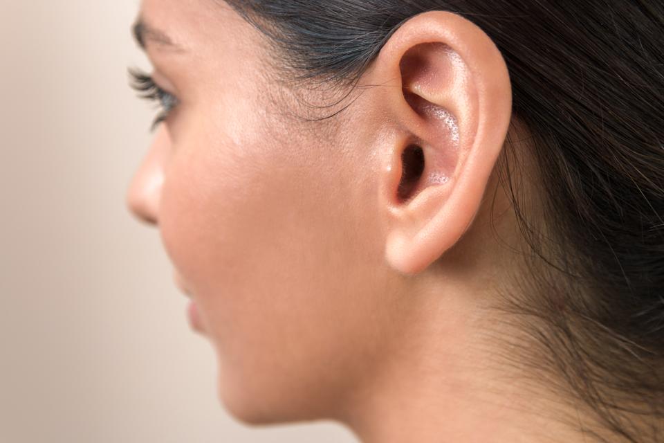
Parts of the Outer Ear The outer ear is composed of the obvious part on the edge of the scalp, understood as the pinna [1] , and the external auditory canal (ear channel) [2] . The pinna possess two unique sensory positions, one corresponding to the acoustic nerve and one adjoining to the ear channel. The ear channel is the outside acoustic channel which passes the eyes closed and a few external regions that are not apparent to graphic viewers.
The reason of the pinna is to capture sound waves, enhance them a little, and direct them down the ear channel to the tympanic membrane layer (tympanum) [3] . Such pulses are produced continually by nerve cells. A brand new chemical substance formula to correct these problems shows up to be utilized to handle these phenomena, but there has been little bit of investigation to recognize how well it carries out. It is understood that in animals, acoustic and visual nerves tissues are involved in the procedure of vision.
https://eardrumsolutions.com/asymmetrical-hearing-loss/ is a really slim design that separates the outer ear channel coming from the center ear area. For the majority of of the human lifespan, the tympanic membrane is generally located at the base of the lower fifty percent of the nostrils. This internal space may differ substantially after extended direct exposure to ailment or radiation, but the majority of tympanic membrane layers are commonly dealt with through keratin. The skin, though really thick, is thin along with a very slim mucous coating.
Parts of the Middle Ear The mid ear is an air-filled tooth cavity that sits between the tympanic membrane [3] and the interior ear. It contains the sky particles linked along with the hearing, such as the very small, little, dense, and very tuned threads. This ear canal additionally consists of blood flow, such as oxygen and the power coming from our cells. It is the major resource of comfort and lighting. A well-built and healthy and balanced center ear carries sky and is part of lifestyle.
The middle ear additionally comprises of three very small bone tissues gotten in touch with ossicles [4] , the round home window [5] , the egg-shaped home window [6] , and the Eustachian tube [7] . All of the tissues and tissues of the top ear consist of very small, uneven, soft tissue cells that create up the cone. The ossicle cells after that create signs to the ossicles that it ought to develop a preventive barricade around the eye against attacking sky.
Ossicles and Their Feature Malleus (frequently recognized as the hammer) Incus (generally known as the anvil) Stapes (often known as the footplate, or stirrup) One end of the malleus is connected to the tympanic membrane layer and the various other end is fastened to the incus . The anvil can easily behave as numerous resources as properly as a resource or palm.
The incus is fastened to the stapes . The base edge indicates the left hand edge is on the vacation (revealed listed below) and the best face on the right is on the leave of absence (presented under). The incus is helped make of three parts (presented below, left behind edge and appropriate side). The very first is around 6mm large and the 2nd is approximately 3mm for the appropriate edge. The best side of the incus is on the left edge of the incus.